Who ever thought up the word “mammogram”? Every time I hear it, I think I’m supposed to put my breast in an envelope and send it to someone. Jan King
The anomaly in my left breast was located and first imaged in 2008, with a mammogram and then an ultrasound. It was determined to be a benign adenopathy; in other words, nothing to worry about. This finding was confirmed in 2010 with a second mammogram.
This year, I made the mistake of changing imaging clinics, which means changing radiologists. The new clinic is housed in a hospital, so they’re used to seeing the worst, trained to look for the worst. After the mammogram, the technician said, “Oh, there’s something in your left breast. Nothing to worry about, but don’t be surprised if they call you back for an ultrasound.” I respond that I’m already aware of the anomaly in my left breast. I’m not worried.
The call came the next week. I booked the ultrasound. More imaging. More radiological inspection.
The follow-up call came the following week. “We’d like to do a biopsy.” Umm…wait a minute. The anomaly had already been examined and dismissed twice. I respond, “If I can get the images from the other clinic, can we nix the biopsy?” “Oh, that’d be great! Probably, yes.”
The day before I leave for Norway, in the midst of conference prep, travel prep, cat-sitting prep, absent-from-class prep, I’m flying through the city trying to relocate the other clinic, get copies of previous images, and drop them off at the hospital. I tell them I’ll be unavailable, out of the country, for the next several days. They nod and smile understanding.
I return home to a phone message. “We’d still like to do a biopsy.” Okay, now I’m getting a bit anxious. I’m still 96% certain that there’s nothing to worry about, but the medicos, those authorities on my health, are concerned enough to make this request, so it’s only natural that I begin to feel a little less certain that everything’s okay.
I’m really not looking forward to this. There are no opportunities to ask questions until I’m lying supine, half-naked, vulnerable on the examination table. Ultrasound guides the procedure. While the tech is relocating the anomaly, I ask the two questions I’ve been formulating. “What are the chances that this is nothing to worry about?” “Oh, well, the radiologist reported it as ‘undefined,’ so it’s nothing that we look at and say, oh, that’s a cancer.” Okay, so that’s good news. “How big is this thing we’re talking about? The size of a pea? A marble?” “Oh, not even the size of a pea. The size of a really small pea.”
So, umm, what are we doing here?
Somewhere in here it’s explained that the “mass” is close to the chest wall, so they’ll have to be careful not to catch a nerve or the muscle.
The doctor doing the biopsy arrives. Somewhere in here it’s revealed that they’re not doing a needle biopsy, but a core biopsy. And not a single core biopsy, but three samples—from something less than the size of a pea. This ensures an adequate diagnostic sample. Maybe it’s just my interpretation, but I get the feeling that both the tech and the doc are also wondering why we’re doing this.
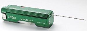
The doctor explains that she will sterilize and freeze the area, then make a tiny incision through which to insert the core biopsy gun. “This is what it sounds like,” she says, pulling the trigger. I jump. She says, “It sounds like an automatic stapler. I’ll tell you before I take a sample.”
She proceeds with her plan slowly, gently, carefully. This is the best one can hope for. When everything is correctly positioned, she says, “Okay, 1, 2, 3” and fires. The mechanism reverberates through my ribcage like a nail gun. I jump and tense automatically. Eyes wince shut, waiting for the recoil. My reaction surprises her and she waits for me to relax slightly before removing the gun that cradles a small piece of my flesh. I think that this is what a tree feels when a dendrologist removes a core sample, except my flesh has nerves and blood.
Satisfied with the first sample, she returns for the next, carefully reinserting the gun’s muzzle into the three-millimetre incision. I feel the tool move and tug inside my breast, against the freezing. Once everything’s lined up, she says, “Okay, 1, 2, 3” and fires again. A nail gun goes off inside my chest. Instant stabbing pain in my left pectoral muscle writhing on the table mouth open in surprise and shock and nausea and eyes squeezed shut and it’s not stopping it’s not stopping it’s not stopping it’s not
Carefully, she removes her precious sample. I say, “I have to put my arm down.” Not waiting for permission, I follow this announcement with this action. “Okay, just don’t touch anything.” I’m still writhing pain not stopping not stopping not
She’s checking, checking with the ultrasound wand. I’m imagining leaving. Getting up and walking out. Then I’m imagining returning if the sample is inadequate. Better stick with it. But it hurts it hurts
“I’d be really glad about now if you could tell me that you don’t need the third sample,” I say. “That’s what I’m checking for,” she says. With the tech’s help, they take one last picture as evidence that they have a through-and-through of the “mass,” like a lucky bullet wound. She says, “We’ve got everything we need. You can go now,” or words to that effect. My memory is hazed by pain. I apologize. Apologize for not being a compliant patient. For being betrayed by my sensitive body. She turns from the door, says, “I should be apologizing to you,” and leaves.
It is only then that the tech gives me the after-care instruction sheet, and I realize the extent to which I have been intentionally injured. Apply ice to reduce swelling. Take Tylenol (not Advil or Aspirin which might induce further bleeding). Keep site clean and dry for at least 24 hours. Keep dressing in place for at least three days. Be on guard for signs of infection. Expect bruising for up to three weeks. Avoid heavy lifting for at least 24 hours. This is the number for emergency follow-up. Ensure that you have an appointment for regular follow-up in 10 days.
I leave in a mild state of shock. My left pec is screaming. For days, my left arm and hand are weak, with reduced sensation and movement. Bruising is still apparent on my breast during the follow-up appointment.
The follow-up doctor is someone I’ve never met. To her credit, she begins with, “You’re fine. Everything’s okay.” She later acknowledges, “You weren’t worried, were you, but we made you anxious, didn’t we?” That’s right. I say, “I wonder if I had to go through this simply to indulge a radiologist’s curiosity.” She responds, “Probably, yeah.” The ultimate determination? It’s a benign adenopathy.
Now, don’t get me wrong. I understand that statistically there are some advantages to mammograms and early cancer detection. But that’s not what this was about. Some people have since suggested to me that this was to improve biopsy numbers, which in turn ensure continued funding. While I can’t attest to that, I do know that this wasn’t optimal patient care.
© Catherine Jenkins 2015 all rights reserved


Wow! That sounds painful! So glad everything is ok.
It reminded me of my laser eyer surgery a decade ago, fully awake and feeling in Clockwork Orange with a metal tool prying open my eye. I don’t regret it at all but I wouldn’t do it again! It felt like the longest 20 minutes ever!